Connective tissue, tendons and remodelling: why mature tendon tissue is barely renewed
Radiocarbon data suggest the Achilles tendon core forms in growth and is barely renewed thereafter: what that means for healing, scar and the equine tendon.
Scientific reviewPosition not yet filled, stated openly.
10 min readLast substantive review
Open access
Executive summary
Tendons consist largely of type I collagen. Radiocarbon measurements suggest that the core of the human Achilles tendon is essentially laid down during longitudinal growth and is barely renewed thereafter. The superficial digital flexor tendon of the horse counts as a functionally comparable energy-storing tendon. Both heal through repair tissue rather than genuine rebuilding: equine tendon scar was found to contain more type III collagen. That fits the high re-injury rates seen in both species.
26primary sources
50 %of them level 1 to 2
4species studied
1980–2026publication years
Key points
Carbon-14 measurements in 28 human Achilles tendons suggest that the tendon core is formed in roughly the first 17 years of life and essentially not replaced thereafter; skeletal muscle from the same bodies showed ongoing turnover.
In an analysis of 10,262 Japanese Thoroughbreds, forelimb tendonitis affected 11.1 per cent of the horses; in the hindlimb it barely occurred at all.
Scar tissue examined from equine tendons contained 20 to 30 per cent type III collagen, normal tendon tissue almost exclusively type I: healing does not replace the tissue, it patches it.
Loading changes a tendon measurably without exchanging its core: across 27 intervention studies with 264 adults, stiffness rose above all, and the magnitude of the load mattered more than the form of exercise.
The shared mechanism is well supported, the clinical transfer is not: treatment results in the horse are no evidence for the human, and vice versa.
What a tendon is made of, and what sets it apart from other connective tissue
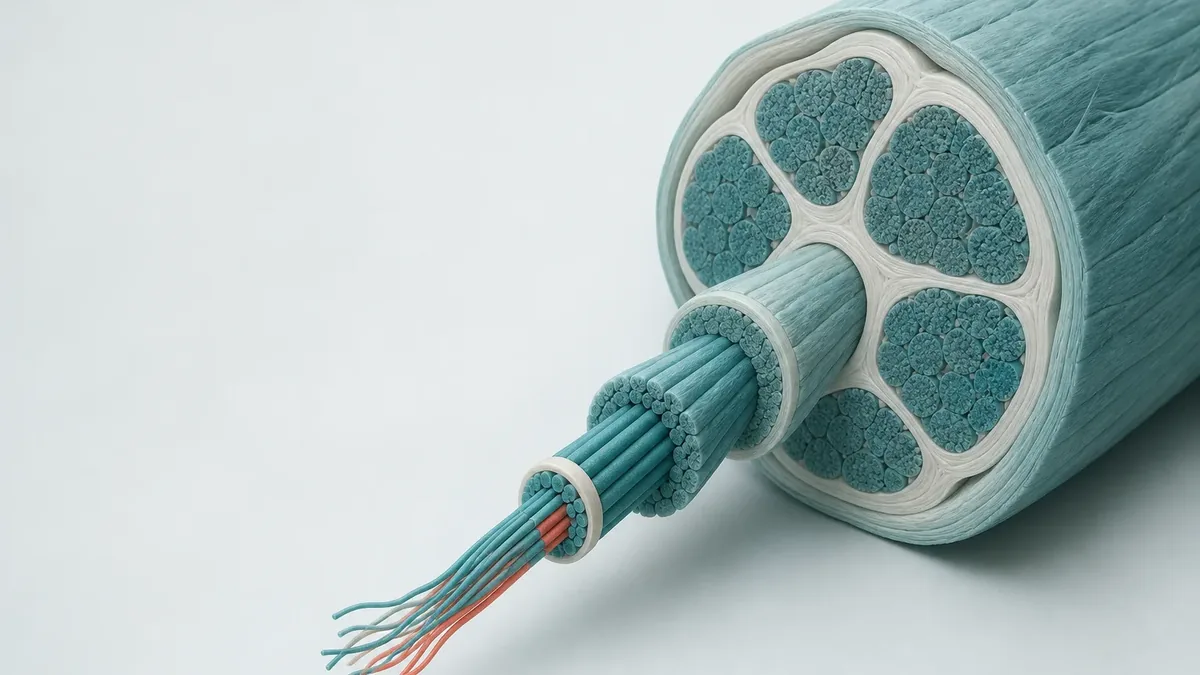
Tendons are connective tissue built for tension. Most of their dry weight is type I collagen, which bundles up from fibrils into fibres and then into fascicles, the largest subunits of a tendon. Between those fascicles sits the interfascicular matrix: a softer layer that is rich in elastin in the equine tendons studied. Mechanical testing on excised equine tendons showed that it is not the fascicles themselves but precisely this intervening layer that decides how far a tendon can stretch and how completely it springs back.
What separates an energy-storing tendon from a positional tendon?
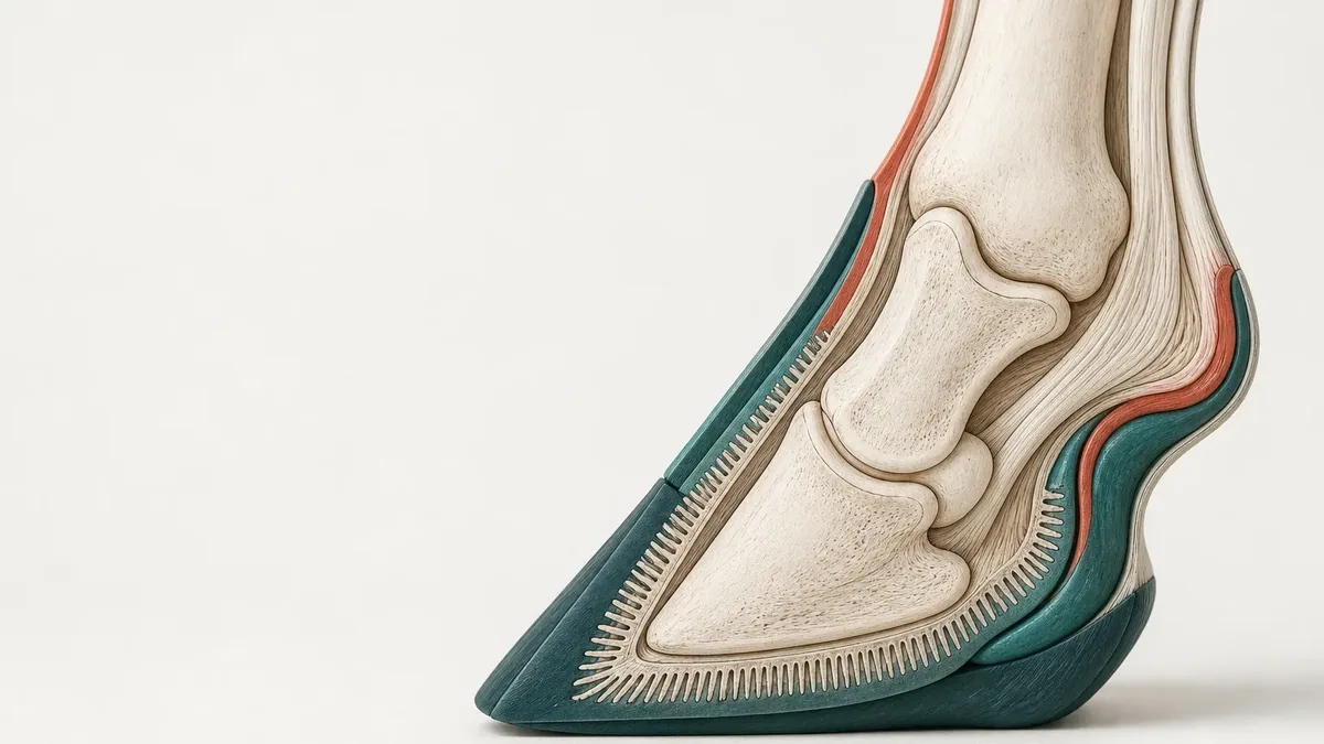
A positional tendon transmits force and sets a joint. An energy-storing tendon does the same, and on top of that takes up elastic energy at foot strike and returns it at push-off. That lowers the energy cost of locomotion, but it demands strains close to the failure load. The human Achilles tendon and the superficial digital flexor tendon of the horse both belong to the second group: same job, same narrow safety margin.
In a direct comparison of two equine tendons with different jobs, the energy-storing tendon was the one with the lower values for collagen building instructions and for degradation markers, despite its higher cell density. The purely positional tendon from the same limb turned its matrix over noticeably faster. The authors consider it possible that this very slowdown in the energy-storing tendon favours the accumulation of damage: the tissue under the greatest strain is the one that renews itself least.
The nuclear-bomb clock: how often a mature tendon is really renewed
How fast tendon tissue turns over was an open question for a long time. Published estimates of the half-life of tendon protein ranged from two months to two hundred years, a difference of more than a thousandfold. A Copenhagen research group settled the question by a detour: the atmospheric nuclear weapons tests between 1955 and 1963 sharply raised the carbon-14 content of the air. Tissue laid down at that time carries the signature of its year of manufacture.
In 28 forensic samples of human Achilles tendon, the carbon-14 content measured matched atmospheric values from several decades before the samples were taken. On that calculation the tendon core is laid down in roughly the first 17 years of life, that is during longitudinal growth, and is essentially not replaced afterwards. Four skeletal muscle samples from the same bodies, by contrast, showed ongoing turnover. In the group's own assessment, the finding probably explains why tendons regenerate so poorly.
Does that mean training does not change the tendon at all?
No. A synthesis of 27 intervention studies with 264 healthy adults in total found clear increases in tendon stiffness after at least eight weeks of raised loading, and smaller increases in cross-sectional area. What mattered was the magnitude of the load, not the type of muscle contraction. A broader analysis of 61 papers with 763 people reached the same result and showed in addition that the rise in stiffness comes mainly from altered material properties and only in small part from a thicker tendon. Core remodelling and measurable adaptation are therefore two different processes: the tendon becomes stiffer and somewhat thicker without its load-bearing scaffold being exchanged.
The same group later compared diseased and healthy tendons. In 25 tendinopathic Achilles tendons a substantial share of the collagen had been renewed in adulthood; in 10 healthy tendons it had not. The modelling suggested that roughly half of the collagen in the diseased tendon had already been slowly replaced years before the first symptoms. Whether this raised turnover is a very early phase of disease or a pre-existing risk factor cannot be decided from these data.
The horse as a model: superficial digital flexor tendon and Achilles tendon
In comparative research, injury to the superficial digital flexor tendon in the sport horse counts as one of the best-supported animal models of a human disorder, namely overstrain injury of the Achilles tendon. A review from 2014 notes that no equivalent rodent or rabbit model exists for this function. The reason is simple: few species move on a tendon that works close to its material limit at the gallop.
The frequency is striking. In an analysis of 10,262 Japanese Thoroughbreds from a single year group, forelimb tendonitis affected 11.1 per cent of the horses, while the same injury in the hindlimb, with six cases, barely occurred at all. A British ultrasound series in 263 National Hunt horses found tendon changes in 24 per cent of the animals over two seasons, with differences between training yards of 10 to 40 per cent that the authors did not, however, class as established.
Why is it almost always the same tendon?
Because function, load and age coincide. The energy-storing tendon of the forelimb carries most of the load at the gallop, its remodelling is slow, and damage adds up over the years. In the Japanese analysis, horses aged three and over were far more affected than two-year-olds. What did not work is just as telling: routine ultrasound at three-month intervals did not predict the acute rupture in the British series.
The interfascicular matrix: where an energy-storing tendon gets its stretch
Pull fascicles from an energy-storing tendon and from a positional tendon in the same equine limb, and the fascicles themselves barely differ. The difference lies in between. The interfascicular matrix of the energy-storing tendon allows more sliding between neighbouring fascicles and springs back more elastically, so it recovers better after repeated loading. With advancing age this layer became stiffer in the energy-storing tendon and tolerated loading cycles less well, while the fascicles remained largely unchanged.
A key reason for that is elastin. Biochemical and immunohistological work localised elastin specifically to the interfascicular matrix of the energy-storing tendon; with age its quantity fell and its arrangement became more irregular. In a further experiment on excised equine tendons, targeted enzymatic degradation of elastin lowered the recovery capacity and the fatigue resistance of exactly that layer, without measurably changing the mechanics of the fascicles.
Healing means scar, not new build
As early as 1980, a study of partially ruptured equine tendons showed the decisive difference: normal tendon tissue consists almost entirely of type I collagen, whereas the scar tissue additionally contained 20 to 30 per cent type III collagen. Type III forms thinner fibrils that bear less load. The repair cells also resembled myofibroblasts more than tendon cells, which suggests that they do not come from the tendon itself but have migrated in from the surrounding tissue.
More recent work supports this picture. In an experiment on isolated tendon fascicles from rat tails, a single strain above two per cent was already enough to degrade the material properties, while structurally visible collagen damage appeared only from six per cent upwards. Over several days in culture, the tendon core repaired that damage barely at all. The authors conclude that the resident cells mainly break tissue down and send signals outwards instead of rebuilding it themselves.
Why does a healed tendon so often rupture again?
Because the repair tissue matches the original neither structurally nor mechanically. A review from 2022 describes the typical failures: inflammation that does not settle in time, excessive ingrowth of blood vessels, and immature tissue that never reaches the mechanical competence required. Without appropriate mechanical stimuli the fibre alignment stays disordered, and the cross-linking that gives a tendon its stiffness forms only incompletely.
What the treatment data show, horse and human side by side
In the horse, implantation of the animal's own bone marrow cells into the injured tendon has been followed for years. In a follow-up of 113 racehorses over at least two years after their return to full work, the re-injury rate was 27.4 per cent, and 25.7 per cent among the National Hunt horses. That was lower than in previously published comparison groups, though without a concurrent control group. A small randomised trial in 12 horses with career-ending injury found a better-ordered fibre structure in the treated tendons after six months.
A systematic analysis from 2024 pooled 21 studies of cell and plasma treatments in the horse. Cell treatments did not raise the rate of return to performance, but they did lower the risk of re-injury. The authors explicitly stress the weak quality of the studies. A comparative review of 123 papers on platelet-rich plasma in horse and human found positive effects mostly in studies at high risk of bias, and absent effects more often in the methodologically better ones.
In humans the evidence base is larger, but no more satisfying. A synthesis of 33 studies with just under 36,000 people found that after acute Achilles tendon rupture, surgery brought fewer re-ruptures and more frequent return to sport, but also more infections and more nerve injuries. An analysis of 14 randomised trials with 1,628 participants showed a similar pattern: fewer re-ruptures after surgery, but more complications after the open procedure. Platelet-rich plasma changed the outcomes in neither treatment pathway.
For the painful, unruptured tendon, loading itself does the work. A randomised trial in 58 people with chronic Achilles tendon complaints compared eccentric training with heavy slow resistance training: both clearly improved function and pain, the effect held at 52 weeks, and no difference between the approaches emerged. An analysis of 21 randomised trials with 994 people supports this picture: against other forms of training, eccentric training was equivalent. For shockwave therapy, by contrast, a review at the Achilles tendon found only small and inconsistent effects, and a network analysis of non-surgical approaches could not name a best option.
Convergence and divergence: what transfers and what does not
The mechanism transfers. Both tendons store elastic energy, both have a core that is remodelled very slowly, both heal through scar tissue, in both the risk rises with age, and in both part of the specialisation sits in the matrix between the fascicles. This pattern is consistent across independent measurement methods and across several species, which makes a shared biology far more likely than coincidence.
The clinical picture as a whole does not transfer. The review from 2014 states explicitly that the equine tendon is a model for the acute rupture in athletes, not for the full spectrum of human tendon complaints and in particular not for chronic tendon pain. The loading profiles also differ fundamentally: a racehorse carries half a tonne on four limbs at the gallop, a human runs upright on two.
What can soberly be drawn from all this
Three observations hold up to scrutiny. First, in the adaptation of healthy tendons the magnitude of loading appears to be the more effective lever than the chosen form of exercise: the synthesis of intervention studies found an association with intensity, not with contraction type, and the head-to-head comparison of two training forms in symptomatic tendons produced no difference. Second, prevention weighs more heavily than repair, because on current knowledge the tendon core is not built anew.
Third, supplements are no substitute for loading. A synthesis of 19 studies with 768 adults found favourable effects on tendon morphology under collagen peptides combined with regular training, but rated its own certainty in that statement as very low. Without a training stimulus, nothing comparable was shown in this body of work.
No procedure so far turns scar tissue back into normal tendon tissue, neither in the horse nor in the human.
No screening reliably predicted acute tendon rupture in the series available.
No treatment result in one species permits a conclusion about the other.
None of the work cited here supports a dosing or treatment recommendation.
Measurement methods for tendon remodelling: what they see and what they systematically miss
Original analysis
Compiled from the primary studies cited in this article: for each method we recorded which quantity was actually measured, what period it gives information about, and which question it leaves open.
Method
Quantity measured
Time horizon
Blind spot
Species in the cited studies
Carbon-14 bomb pulse
Year of formation of the collagen in the tendon core
Lifelong, decades
Says nothing about the current synthesis rate
Human
Messenger RNA for collagen I and III
Current building instructions of the cells
Hours to days
No evidence that the protein is actually incorporated
Horse
Degradation markers of type I collagen
Ongoing breakdown of the existing matrix
Days to weeks
Does not separate breakdown from new formation
Horse
Tissue autofluorescence
Non-enzymatic cross-linking, indirectly matrix age
Years
Non-specific as to the cause
Horse
Collagen hybridising peptide
Mechanically unfolded collagen triple helix
Immediately after loading
Does not capture functional damage below the detection threshold
Rat, outside the body
Ultrasound cross-sectional area
Size and echo pattern of the tendon
Weeks to months
Did not predict the acute rupture in a two-year series
Horse
Stiffness measurement in the living
Mechanical response of the tendon to force
Weeks to months
Separates material change from shape change only indirectly
Human
Limitations and uncertainty
The central data series on human tendon turnover rests on forensic samples and a back-calculation from atmospheric values, not on repeated measurements in the same living people.
On the biology of tendon remodelling there are almost only laboratory, tissue and observational data; randomised trials exist mainly on treatment. About half of the work used here is a systematic review, meta-analysis or randomised trial, the other half is mechanistic or observational. A higher proportion is not currently available in this field.
The equine cell treatment data rest in part on comparisons with historical control groups; the systematic analysis from 2024 rates the study quality as weak and the risk of bias as high.
The prevalence figures come from Thoroughbreds under racing conditions in Japan and Great Britain and cannot be transferred to leisure horses, to other disciplines or to other countries.
The findings on the interfascicular matrix and on elastin come from experiments on excised tissue; a clinical link with injury risk in the living animal is therefore not proven.
On the effect of collagen peptides on tendon tissue, the available synthesis rates the certainty of its own statement as very low.
Open questions
Is the raised collagen turnover that begins years before the first symptoms a very early phase of disease, or a pre-existing risk factor?
Can the state of the interfascicular matrix be measured in the living animal or human before the tendon ruptures?
Is there a form of loading that actually lowers the proportion of type III collagen in repair tissue and normalises the fibre alignment?
Why do cell treatments in the horse lower the re-injury rate without raising the return to full performance?
Frequently asked questions
Can damaged tendon tissue be built back up?
On current knowledge, not in the sense of a genuine new build. The load-bearing core of the human Achilles tendon is laid down during longitudinal growth and is essentially not replaced afterwards. After an injury, repair tissue forms; in equine tendons it contains 20 to 30 per cent type III collagen, whereas healthy tendon tissue consists almost entirely of type I. This scar tissue bears less load. Loading can make a tendon stiffer and somewhat thicker, but it does not exchange the scaffold.
Why does a tendon heal so much more slowly than a muscle?
Because the two tissues handle their matrix in fundamentally different ways. In the same bodies in which the tendon core held collagen decades old, skeletal muscle showed ongoing turnover. Tendon tissue is also poor in cells and poorly supplied with blood. In a laboratory experiment on fascicles from rat tail tendons, mechanical damage went essentially unrepaired over several days: the resident cells tended to break tissue down and send signals outwards instead of rebuilding it themselves.
Do collagen supplements do anything for the tendons?
The evidence is weak and the conclusion uncertain. A synthesis of 19 studies with 768 healthy adults found favourable effects on tendon morphology under collagen peptides combined with regular training. The authors themselves, however, rated their certainty in exactly that statement as very low. Without accompanying training, nothing comparable was shown. This text deliberately names no amounts and gives no recommendation.
Is the flexor tendon of the horse really the same as my Achilles tendon?
Functionally very similar, clinically not identical. Both are energy stores that take up energy at foot strike and give it back at push-off, both work close to their material limit, both have a slowly remodelled core and heal through scar. A review from 2014 states explicitly, however, that the equine tendon is a model for the acute rupture in athletes, not for the whole spectrum of human tendon complaints and in particular not for chronic tendon pain.
Why do older horses and older people injure their tendons more often?
Because damage accumulates and the matrix between the fascicles ages. In an analysis of 10,262 Japanese Thoroughbreds, horses aged three and over had a markedly higher risk than two-year-olds. At tissue level, the interfascicular matrix of the energy-storing tendon became stiffer and more prone to fatigue with age, and its elastin decreased and arranged itself more irregularly. That explains the link plausibly, but it is a laboratory finding and not an individual risk marker.
Does an Achilles tendon rupture have to be operated on?
That question is decided by the treating doctor, not by an article. The evidence shows a trade-off: a synthesis of 33 studies with just under 36,000 people found fewer re-ruptures and more frequent return to sport after surgery, but also more infections and more nerve injuries. An analysis of 14 randomised trials with 1,628 participants likewise showed fewer re-ruptures after surgery, but more complications after the open procedure. Platelet-rich plasma improved the outcomes in neither group.
Sources
Heinemeier KM, Schjerling P, Heinemeier J, Magnusson SP, Kjaer M. Lack of tissue renewal in human adult Achilles tendon is revealed by nuclear bomb 14C. FASEB Journal, 2013 (Laboratory study | Human)DOI 10.1096/fj.12-225599 Carbon-14 measurements in 28 Achilles tendons and 4 muscle samples showed that the tendon core is essentially formed in roughly the first 17 years of life and barely renewed thereafter, while muscle turns over continuously.
Heinemeier KM, Schjerling P, Ohlenschlaeger TF, Eismark C, Olsen J, Kjaer M. Carbon-14 bomb pulse dating shows that tendinopathy is preceded by years of abnormally high collagen turnover. FASEB Journal, 2018 (Case-control study | Human)DOI 10.1096/fj.201701569R In 25 tendinopathic versus 10 healthy Achilles tendons, a substantial share of the collagen had been renewed in adulthood; the modelling points to years of raised turnover before symptom onset.
Patterson-Kane JC, Rich T. Achilles tendon injuries in elite athletes: lessons in pathophysiology from their equine counterparts. ILAR Journal, 2014 (Other | Multiple species)DOI 10.1093/ilar/ilu004 The review rates the superficial digital flexor tendon of the horse as a functionally and clinically equivalent model of the human Achilles tendon, but stresses that it represents only the acute rupture and not chronic tendon pain.
Birch HL, Worboys S, Eissa S, Jackson B, Strassburg S, Clegg PD. Matrix metabolism rate differs in functionally distinct tendons. Matrix Biology, 2008 (Laboratory study | Horse)DOI 10.1016/j.matbio.2007.10.004 Despite a higher cell density, the energy-storing flexor tendon showed lower collagen messenger RNA, lower degradation markers and higher tissue fluorescence than the positional tendon, which points to slower matrix remodelling.
Thorpe CT, Godinho MSC, Riley GP, Birch HL, Clegg PD, Screen HRC. The interfascicular matrix enables fascicle sliding and recovery in tendon, and behaves more elastically in energy storing tendons. Journal of the Mechanical Behavior of Biomedical Materials, 2015 (Laboratory study | Horse)DOI 10.1016/j.jmbbm.2015.04.009 The interfascicular matrix of the energy-storing tendon behaved more elastically than that of the positional tendon, and became stiffer and less resistant to repeated loading with advancing age.
Godinho MSC, Thorpe CT, Greenwald SE, Screen HRC. Elastin is localised to the interfascicular matrix of energy storing tendons and becomes increasingly disorganised with ageing. Scientific Reports, 2017 (Laboratory study | Horse)DOI 10.1038/s41598-017-09995-4 Elastin is concentrated in the interfascicular matrix of energy-storing equine tendons; quantity and order decline with age, which is consistent with the raised injury risk of old energy-storing tendons.
Kasashima Y, Takahashi T, Smith RKW, Goodship AE, Kuwano A, Ueno T, Hirano S. Prevalence of superficial digital flexor tendonitis and suspensory desmitis in Japanese Thoroughbred flat racehorses in 1999. Equine Veterinary Journal, 2004 (Cross-sectional study | Horse)DOI 10.2746/0425164044890580 In 10,262 Thoroughbreds the prevalence of forelimb flexor tendonitis was 11.1 per cent and hindlimb 0.06 per cent, with a markedly higher risk from the third year of life onwards.
Avella CS, Ely ER, Verheyen KLP, Price JS, Wood JLN, Smith RKW. Ultrasonographic assessment of the superficial digital flexor tendons of National Hunt racehorses in training over two racing seasons. Equine Veterinary Journal, 2009 (Cohort study | Horse)DOI 10.2746/042516409x391042 In 263 National Hunt horses a tendon change was found in 24 per cent of cases over two seasons, with a spread between training yards of 10 to 40 per cent that was classed as not established; routine ultrasound at three-month intervals could not predict the acute rupture.
Williams IF, Heaton A, McCullagh KG. Cell morphology and collagen types in equine tendon scar. Research in Veterinary Science, 1980 (Laboratory study | Horse)PMID 7414083 Normal equine tendon tissue consisted almost entirely of type I collagen; the scar tissue after partial rupture additionally contained 20 to 30 per cent type III, which implies lower tensile strength of the scar.
Stauber T, Blache U, Snedeker JG. Tendon tissue microdamage and the limits of intrinsic repair. Matrix Biology, 2019 (Laboratory study | Rodent)DOI 10.1016/j.matbio.2019.07.008 Isolated tendon fascicles lost elastic modulus from two per cent strain onwards, structural collagen damage appeared from six per cent, and the tendon core repaired that damage barely at all over several days.
Schulze-Tanzil GG, Delgado-Calcares M, Stange R, Wildemann B, Docheva D. Tendon healing: a concise review on cellular and molecular mechanisms with a particular focus on the Achilles tendon. Bone & Joint Research, 2022 (Other | Multiple species)DOI 10.1302/2046-3758.118.BJR-2021-0576.R1 The review describes persistent inflammation, excessive vascular ingrowth and immature repair tissue as the central reasons why healed tendon tissue does not reach the mechanical competence of the original.
Bohm S, Mersmann F, Arampatzis A. Human tendon adaptation in response to mechanical loading: a systematic review and meta-analysis of exercise intervention studies on healthy adults. Sports Medicine - Open, 2015 (Meta-analysis | Human)DOI 10.1186/s40798-015-0009-9 Across 27 studies and 37 interventions with 264 healthy adults, tendon stiffness and elastic modulus rose clearly and cross-sectional area less so; loading intensity was decisive, contraction type was not.
Beyer R, Kongsgaard M, Hougs Kjaer B, Ohlenschlaeger T, Kjaer M, Magnusson SP. Heavy Slow Resistance Versus Eccentric Training as Treatment for Achilles Tendinopathy: A Randomized Controlled Trial. The American Journal of Sports Medicine, 2015 (Randomised trial | Human)DOI 10.1177/0363546515584760 In 58 people with chronic Achilles tendinopathy, eccentric training and heavy slow resistance training improved function and pain equally and durably out to 52 weeks, with no difference between the approaches.
Yuan F, Ren K, Zhao Z, Wang P, Song L. The efficacy of eccentric exercise in the treatment of Achilles tendinopathy: a systematic review and meta-analysis. BMC Musculoskeletal Disorders, 2026 (Meta-analysis | Human)DOI 10.1186/s12891-026-09861-3 Across 21 randomised trials with 994 people, eccentric training was superior to physical modalities for pain reduction but equivalent to other forms of training; the functional data were highly heterogeneous.
Yang Z, Ge Z, Zheng J, Zhang L, Yang Z. Surgical treatment versus conservative management for acute Achilles tendon rupture: a systematic review and meta-analysis. Journal of Orthopaedic Surgery and Research, 2025 (Meta-analysis | Human)DOI 10.1186/s13018-025-05990-y Across 33 studies with 35,896 people, surgery lowered the re-rupture rate and raised return to sport, but came with more infections and nerve injuries; platelet-rich plasma showed no added benefit.
Xu S, Xiao J, Li Y, Liu E, Qin Y, Shen X. Operative versus nonoperative management of acute Achilles tendon rupture: a systematic review and meta-analysis of clinical outcomes from randomized controlled trials. Annals of Medicine, 2025 (Meta-analysis | Human)DOI 10.1080/07853890.2025.2537349 Across 14 randomised trials with 1,628 participants, open and minimally invasive surgery clearly lowered the re-rupture rate, with a higher complication rate for the open technique and no difference in functional scores.
Ko VM, Cao M, Qiu J, Fong IC, Fu SC, Yung PS, Ling SK. Comparative short-term effectiveness of non-surgical treatments for insertional Achilles tendinopathy: a systematic review and network meta-analysis. BMC Musculoskeletal Disorders, 2023 (Meta-analysis | Human)DOI 10.1186/s12891-023-06170-x Across 9 studies with 464 participants, eccentric training with soft tissue treatment ranked best in the short term, yet confidence in all non-surgical approaches was very low and no best option could be named.
Charles R, Fang L, Zhu R, Wang J. The effectiveness of shockwave therapy on patellar tendinopathy, Achilles tendinopathy, and plantar fasciitis: a systematic review and meta-analysis. Frontiers in Immunology, 2023 (Meta-analysis | Human)DOI 10.3389/fimmu.2023.1193835 For the Achilles tendon the analysis found only small and inconsistent effects of shockwave therapy on pain and function, while it showed clear effects in plantar fasciitis.
Godwin EE, Young NJ, Dudhia J, Beamish IC, Smith RKW. Implantation of bone marrow-derived mesenchymal stem cells demonstrates improved outcome in horses with overstrain injury of the superficial digital flexor tendon. Equine Veterinary Journal, 2012 (Cohort study | Horse)DOI 10.1111/j.2042-3306.2011.00363.x In 113 racehorses followed up, the re-injury rate after cell implantation was 27.4 per cent, and 25.7 per cent in National Hunt horses, which was lower than in published historical comparison groups.
Smith RKW, Werling NJ, Dakin SG, Alam R, Goodship AE, Dudhia J. Beneficial effects of autologous bone marrow-derived mesenchymal stem cells in naturally occurring tendinopathy. PLoS ONE, 2013 (Randomised trial | Horse)DOI 10.1371/journal.pone.0075697 In a randomised trial in 12 horses with career-ending flexor tendon injury, the treated tendons showed a better-ordered structure and a more normal composition than the control tendons after six months.
M'Cloud WRC, Guzman KE, Panek CL, Colbath AC. Stem cells and platelet-rich plasma for the treatment of naturally occurring equine tendon and ligament injuries: a systematic review and meta-analysis. Journal of the American Veterinary Medical Association, 2024 (Meta-analysis | Horse)DOI 10.2460/javma.23.12.0723 Across 21 included studies, cell or plasma treatments did not raise return to performance but did lower the risk of re-injury; study quality was weak and the risk of bias high.
Brossi PM, Moreira JJ, Machado TSL, Baccarin RYA. Platelet-rich plasma in orthopedic therapy: a comparative systematic review of clinical and experimental data in equine and human musculoskeletal lesions. BMC Veterinary Research, 2015 (Systematic review | Multiple species)DOI 10.1186/s12917-015-0403-z Across 123 papers in horse and human, favourable results for platelet-rich plasma occurred mostly in studies at high risk of bias, whereas absent effects were more common in methodologically better studies.
Caspers MK, Gier CJ, Reesink HL. Equine non-septic tenosynovitis: A systematic literature review of site-specific pathological lesions, outcomes and surgical complications. Equine Veterinary Journal, 2024 (Systematic review | Horse)DOI 10.1111/evj.14000 Across 85 studies with 2,101 horses the digital flexor tendon sheath was most often affected, and the probability of returning to the previous level of performance was around 50 per cent for most structures.
Bischof K, Moitzi AM, Stafilidis S, Koenig D. Impact of Collagen Peptide Supplementation in Combination with Long-Term Physical Training on Strength, Musculotendinous Remodeling, Functional Recovery, and Body Composition in Healthy Adults: A Systematic Review with Meta-analysis. Sports Medicine, 2024 (Meta-analysis | Human)DOI 10.1007/s40279-024-02079-0 Across 19 studies with 768 adults, collagen peptides combined with training showed favourable effects on tendon morphology and strength, yet the certainty of the tendon statement was itself rated as very low.
Godinho MS, Thorpe CT, Greenwald SE, Screen HRC. Elastase treatment of tendon specifically impacts the mechanical properties of the interfascicular matrix. Acta Biomaterialia, 2021 (Laboratory study | Horse)DOI 10.1016/j.actbio.2021.01.030 In excised superficial digital flexor tendons from young and old horses, enzymatic degradation of elastin specifically lowered the viscoelasticity, fatigue resistance and recovery capacity of the interfascicular matrix, without measurably changing the mechanics of the fascicles.
Lazarczuk SL, Maniar N, Opar DA, Duhig SJ, Shield A, Barrett RS, Bourne MN. Mechanical, Material and Morphological Adaptations of Healthy Lower Limb Tendons to Mechanical Loading: A Systematic Review and Meta-Analysis. Sports Medicine, 2022 (Meta-analysis | Human)DOI 10.1007/s40279-022-01695-y Across 61 papers with 763 people, mechanical loading clearly raised stiffness and elastic modulus and cross-sectional area only slightly; the stiffness gain came mainly from the material, and protocols with high local tendon strain worked more strongly than those with low strain.